 |
 |
 |
 |
 |
 |
 |
 |
 |
 |

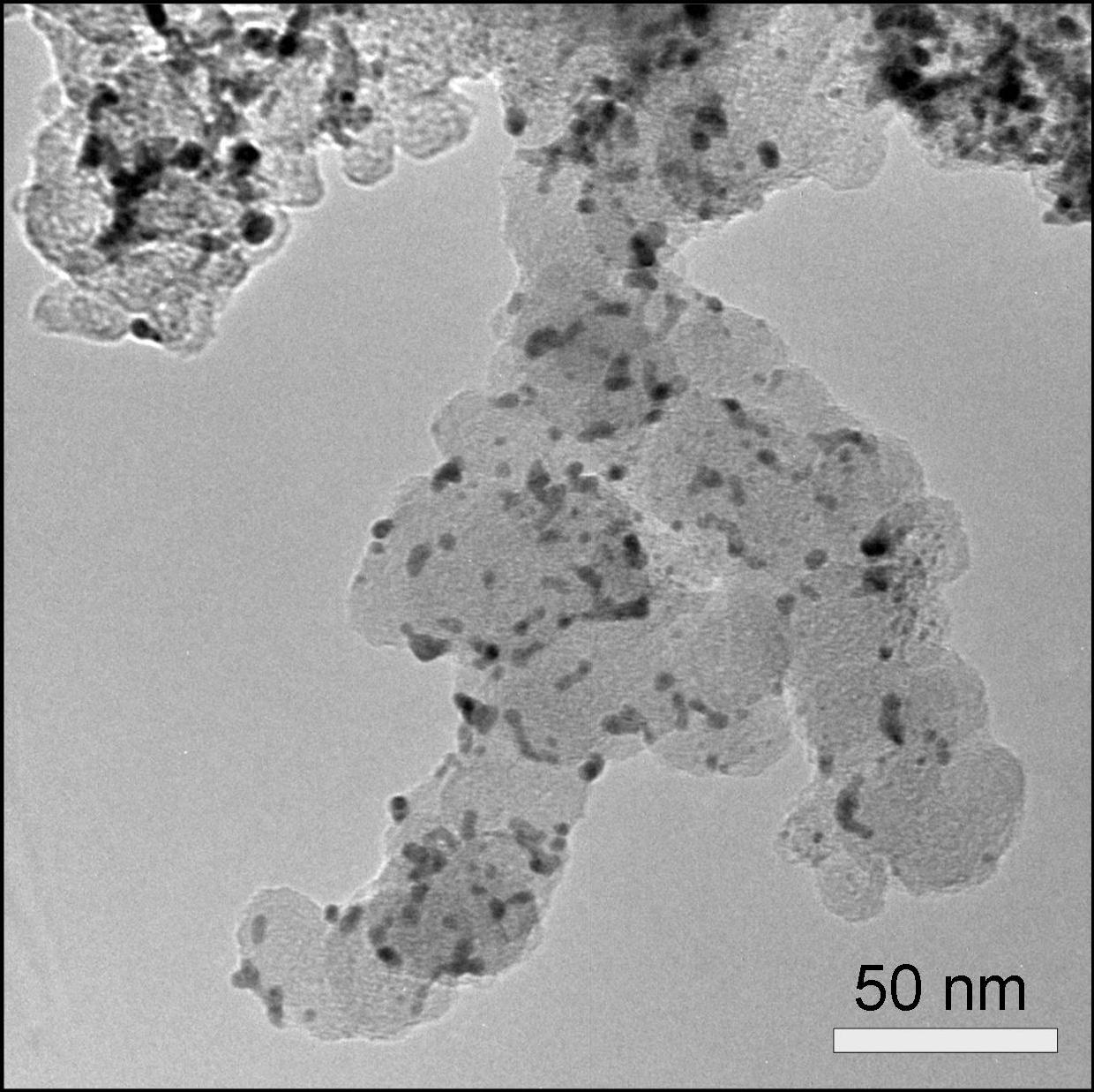
Transmission Electron Microscopy Transmission electron microscopy is a microscopy technique with a significantly higher resolution than light microscopy due to the small de Broglie wavelength of electrons which are used for imaging in a transmission electron microscope. This allows the examination of extremely fine details, e.g. the imaging of a single column of atoms, which is tens of thousands times smaller than the smallest resolvable object in a light microscope. An electron source at the top of the microscope emits electrons that travel through vacuum in the column of the microscope. In a transmission electron microscope, electromagnetic lenses are used to focus and manipulate the electron beam. The electrons interact with the extremely thin specimen (thickness typically below 100 nm) as they pass through. Based on this interaction, an image is formed, subsequently magnified and focused onto a fluorescent screen or onto a CCD camera. Transmission electron microscopy is an ideal tool for the investigation of catalyst material in PEM fuel cells as it can resolve and image the nano-catalyst particles (typical diameters 2-5 nm). One example of a photo of Pt based catalyst particles supported on carbon black taken by transmission electron microscopy is shown below. The small darker spots represent the Pt nanoparticles. The larger, a little brighter, connected sphere-like structures represent the carbon black particles that support the Pt catalyst. This type of photos can be evaluated to generate a catalyst particle diameter distribution which then can be compared with average diameters determined from X-ray diffractometry. | |||||
| |||||