 |
 |
 |
 |
 |
 |
 |
 |
 |
 |

X-Ray Computed Tomography X-ray Computed Tomography (CT) is a nondestructive technique for visualizing the 3-D structures of solid objects which includes also interior structures. This 3D structure is reconstructed from a large series of two-dimensional X-ray images taken at different rotation angles of the sample. The typical spatial resolution of state of the art CT systems is below 1 um. The gray levels in the original 2D CT image correspond to X-ray attenuation, which reflects the proportion of X-rays scattered or absorbed as they pass through they the sample. X-ray attenuation and therefore contrast in the 2D CT images - is primarily a function of the density and composition of the sample material. X-ray computed tomography requires little or no sample preparation and is also very suitable for the investigation of PEM fuel cell components.
|
| ||||
|
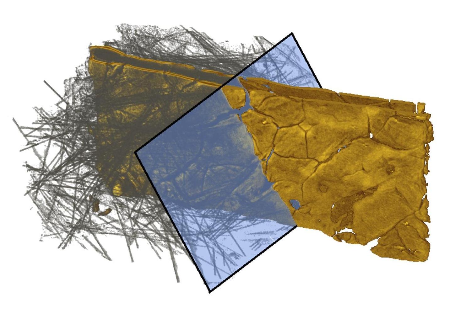
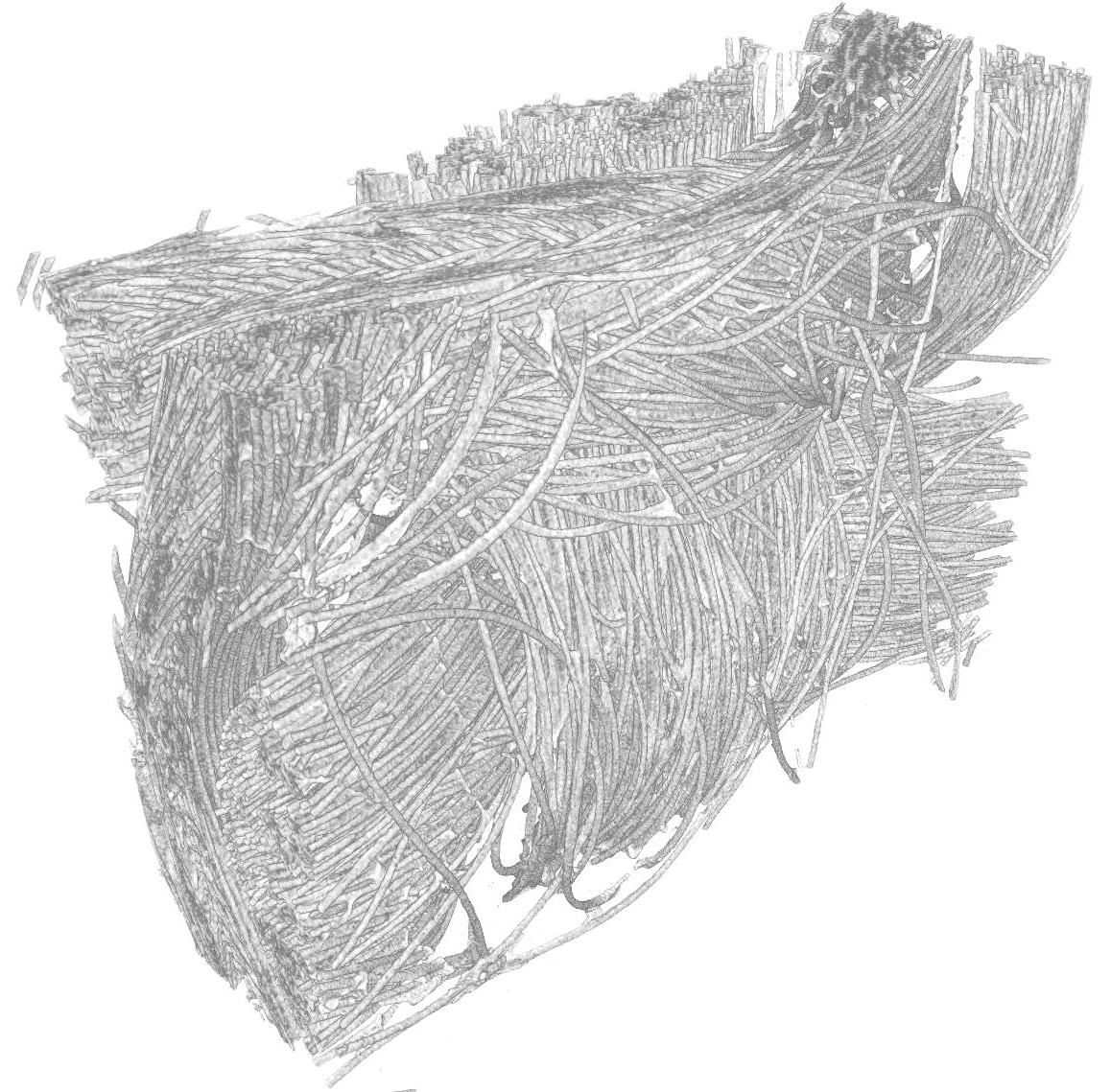
The photo top right shows the 3D structure of a membrane electrode assembly (i.e. membrane, catalyst layers and gas diffusion layers) of a PEM fuel cell as imaged by X-ray computed tomography. The right part of the 3D dataset was manipulated to show only the anode and cathode catalyst layers (in yellow). Cracks are visible within these layers. The photo on the left shows the 3D CT structure of a carbon cloth based gas diffusion layer. The carbon fibers with a diameter of ca. 7 um are cleary resolved and visible (Further CT data see 'Gas Diffusion Layers'). Images published in: A. Pfrang, D. Veyret, G. Tsotridis, G. Janssen, X-ray computed tomography of PEM fuel cells, Proceedings of the International Conference on Materials for Energy 2010, Karlsruhe, Germany
|